sonic svg logo

The Technology Behind VELACUR™

We designed VELACUR as non-invasive test for liver stiffness and attenuation that is accurate, affordable and available at the point of care. AI Guidance and S-WAVE technology go further to set VELACUR apart from alternative solutions. Here’s how.

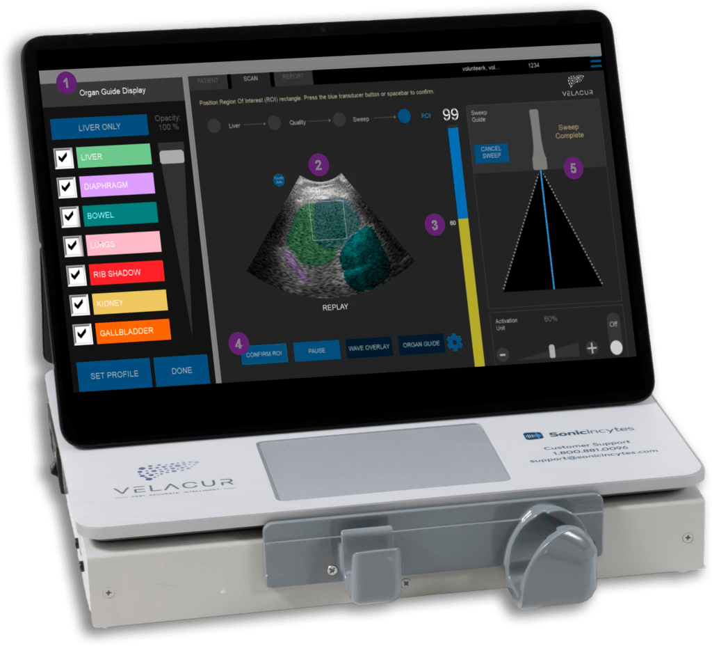
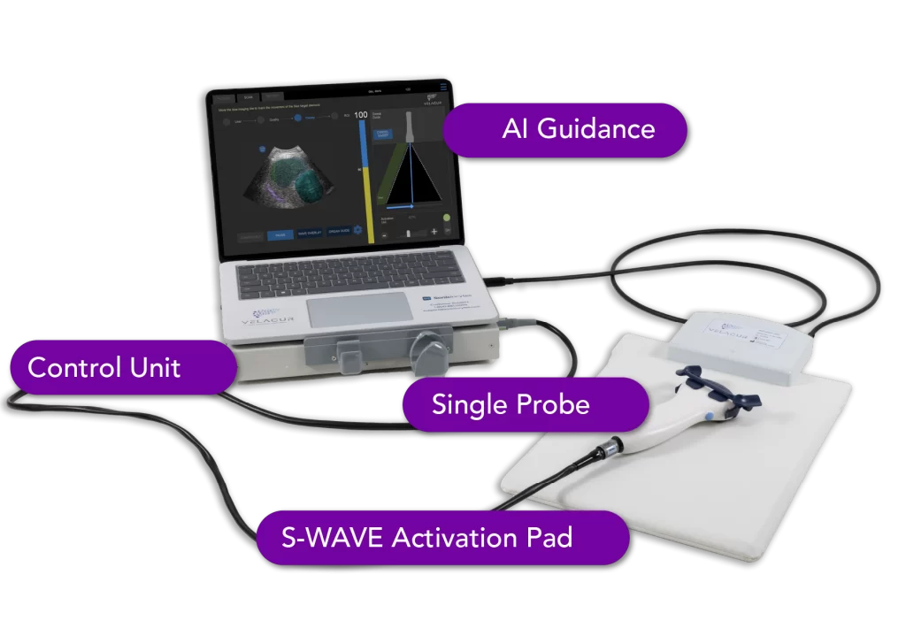
Full touch screen with pre-installed AI-guided software.

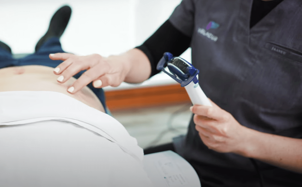
Like MRE, Velacur uses an external activation source to generate shear waves. 

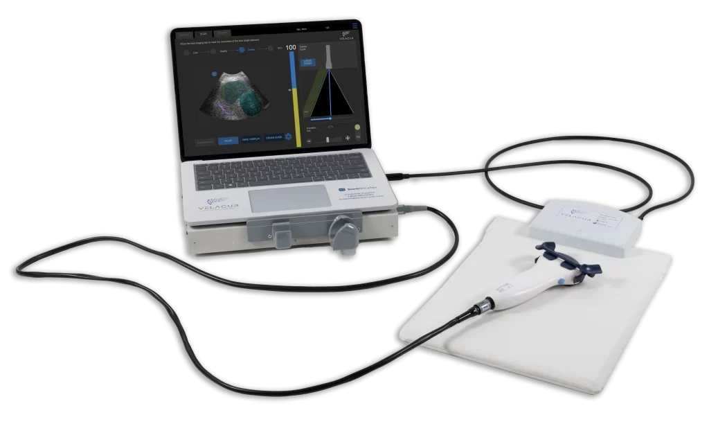
Connects all parts of the system together.

A single probe for a range of body types (BMIs of 25-45)
that does not require re-calibration. 

VELACUR uses artificial intelligence to guide operators in real time,  facilitating liver identification and optimizing wave quality. Making it easy for operators to get measurements they’re confident in, regardless of their sonography experience. 

S-WAVE technology and our advanced probe work together to increase measurement depth and collect a larger sample volume, with 2X the depth and 30X the volume of transient elastography. Our external activation source, similar to MRE, ensures shear wave penetration throughout the abdomen, even in high BMI patients.

Measure confidently with AI Guidance

VELACUR uses artificial intelligence to guide operators in real time, making it easy for operators to get measurements they’re confident in, regardless of their sonography experience. 

Velacur Organ Guide on b-mode image

Organ Guide: color-coded B-Mode overlay. Toggle the overlay on and off with the side menu below. 

B-Mode Imaging for Liver Visualization

Wave Quality Detector: overlay displays wave presence in the visualized area. 

Region of Interest: area where elasticity and attenuation are quantified.

Sweep Guide: supports operators to use the probe in a sweeping motion

Easily locate the liver with our Organ Guide

The Organ Guide overlays the B-Mode image, identifying and color-coding the organs and surrounding anatomy, making it easy for operators to locate the liver. This optional overlay supports operators without sonography experience to use VELACUR confidently.

Optimize measurements with the Wave Quality Detector

The Wave Quality Detector monitors and confirms the presence of good-quality waves. The vertical quality bar provides real-time feedback on the quality of waves during a sweep. This optional overlay enables operators to optimize the quality of waves in the visualized area. 

Why S-WAVE Technology

Deeper measurements

VELACUR boasts a measurement depth of up to 15 cm, 2X deeper than transient elastography. Our single probe is suitable for BMI’s of 25 – 45 with no calibration required, for easy use with a range of patients.

Larger tissue samples

The VELACUR probe is used in a sweeping motion to generate a 3D tissue sample of up to 100cm3 to measure attenuation. Our attenuation coefficient, ACE, is calculated with a tissue sample that is 5000 times larger than a liver biopsy and 30X larger than transient elastography.

External shear wave generation

Based on established ultrasound elastography principles, VELACUR sets itself apart using an external vibration source, similar to Magnetic Resonance Elastography (MRE). The external vibrations are generated through the activation pad, which is placed under the patient’s abdomen, ensuring the presence of shear waves throughout the abdomen, even in high-BMI patients.

Multi-frequency signal

Multiple wave frequencies are used to safeguard against potential measurement errors that could occur with a single frequency. The amplitude, or strength, of the waves can easily be adjusted.

See VELACUR™ in Action

With VELACUR there’s now a practical office-based solution to assess liver tissue stiffness and attenuation in patients with chronic liver disease. The procedure is quick, comfortable and non-invasive and can be performed by any trained medical professional.

person conducting ultrasound imaging on abdomen of patient
Play Video about person conducting ultrasound imaging on abdomen of patient

The Technology Behind VELACUR™

VELACUR uses artificial intelligence to guide operators in real time,  facilitating liver visualization and optimizing wave quality. Making it easy for operators to get measurements they’re confident in, regardless of their sonography experience.

S-WAVE technology and our advanced probe work together to increase measurement depth and collect a larger sample volume, with 2X the depth and 30X the volume of transient elastography. Our external activation source, similar to MRE, ensures shear wave penetration throughout the abdomen, even in high BMI patients.

Measure confidently with AI Guidance

VELACUR uses artificial intelligence to guide operators in real time, making it easy for operators to get measurements they’re confident in, regardless of their sonography experience.

Velacur Organ Guide on b-mode image

Easily locate the liver with the Organ Guide

The Organ Guide overlays the B-Mode image, identifying and color-coding the organs and surrounding anatomy, making it easy for operators to locate the liver. This optional overlay supports operators without sonography experience to use VELACUR confidently.

Optimize measurements with the Wave Quality Detector

The Wave Quality Detector monitors and confirms the presence of good-quality waves. The vertical quality bar provides real-time feedback on the quality of waves during a sweep. This optional overlay enables operators to optimize the quality of waves in the visualized area. 

Why S-WAVE technology

Deeper measurements

VELACUR boasts a measurement depth of up to 15 cm, 2X deeper than transient elastography. Our single probe is suitable for BMI’s of 25 – 45 with no calibration required, for easy use with a range of patients.

Larger, 3D tissue samples

The VELACUR probe is used in a sweeping motion to generate a 3D tissue sample of up to 100cm3 to measure attenuation. Our attenuation coefficient, ACE, is calculated with a tissue sample that is 5000 times larger than a liver biopsy and 30X larger than transient elastography.

External shear wave generation

Based on established ultrasound elastography principles, VELACUR sets itself apart using an external vibration source, similar to Magnetic Resonance Elastography (MRE). The external vibrations are generated through the activation pad, which is placed under the patient’s abdomen, ensuring the presence of shear waves throughout the abdomen, even in high-BMI patients.

Multi-Frequency Signal

Multiple wave frequencies are used to safeguard against potential measurement errors that could occur with a single frequency. The amplitude, or strength, of the waves can easily be adjusted.

See VELACUR™ in Action

person conducting ultrasound imaging on abdomen of patient
Play Video about person conducting ultrasound imaging on abdomen of patient

With VELACUR there’s now a practical office-based solution to assess liver tissue stiffness and attenuation in patients with chronic liver disease. The procedure is quick, comfortable and non-invasive and can be performed by any trained medical professional.